查看更多
密码过期或已经不安全,请修改密码
修改密码
壹生身份认证协议书
同意
拒绝

同意
拒绝

同意
不同意并跳过


Stroke & Vascular Neurology(SVN)最新上线文章“Somatic GJA4 mutation in intracranial extra-axial cavernous hemangiomas”,来自来自首都医科大学附属北京天坛医院、国家神经系统疾病临床医学研究中心、北京神经外科研究所曹勇教授团队等。
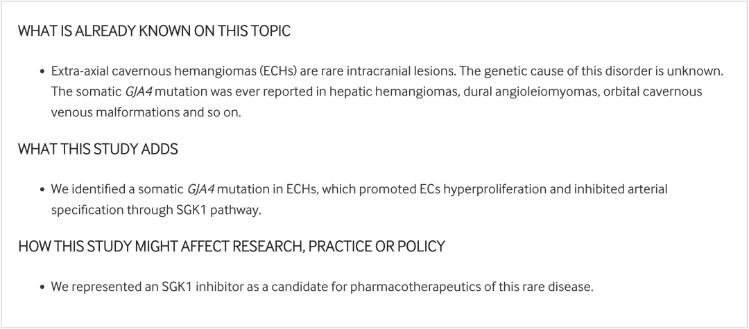
颅内轴外海绵状血管瘤(Extra-axial cavernous hemangiomas, ECHs)是一种偶发性、罕见的颅内占位性病变,多发生于海绵窦内,其病因仍未可知。
研究对12例患者(实验队列)的ECH病变进行了全外显子组测序,并利用液滴数字聚合酶链反应(droplet digital polymerase-chain-reaction, ddPCR)来确认另外46例患者(验证队列)中已识别的突变,进行激光捕获显微切割(Laser capture microdissection, LCM)以捕获和和表征组织细胞亚群。在人脐静脉内皮细胞和新建立的小鼠模型中进行了机制和功能研究。
研究结果显示,实验队列中的5/12例ECH患者中检测到体细胞GJA4突变(c.121G>T, p.G41C),并在验证队列中证实了这一发现(16/46)。LCM和ddPCR相继显示,该突变在病变内皮中富集。内皮细胞的体外实验表明,GJA4突变激活了SGK-1信号,进而上调了参与细胞过度增殖和动脉特征丧失的关键基因。与野生型同窝小鼠相比,过表达GJA4的突变小鼠在出生后3周内于视网膜浅表血管丛中出现了类似ECH样病理形态学特征(静脉管腔扩张和血管密度升高),这些特征被SGK1抑制剂EMD638683逆转。
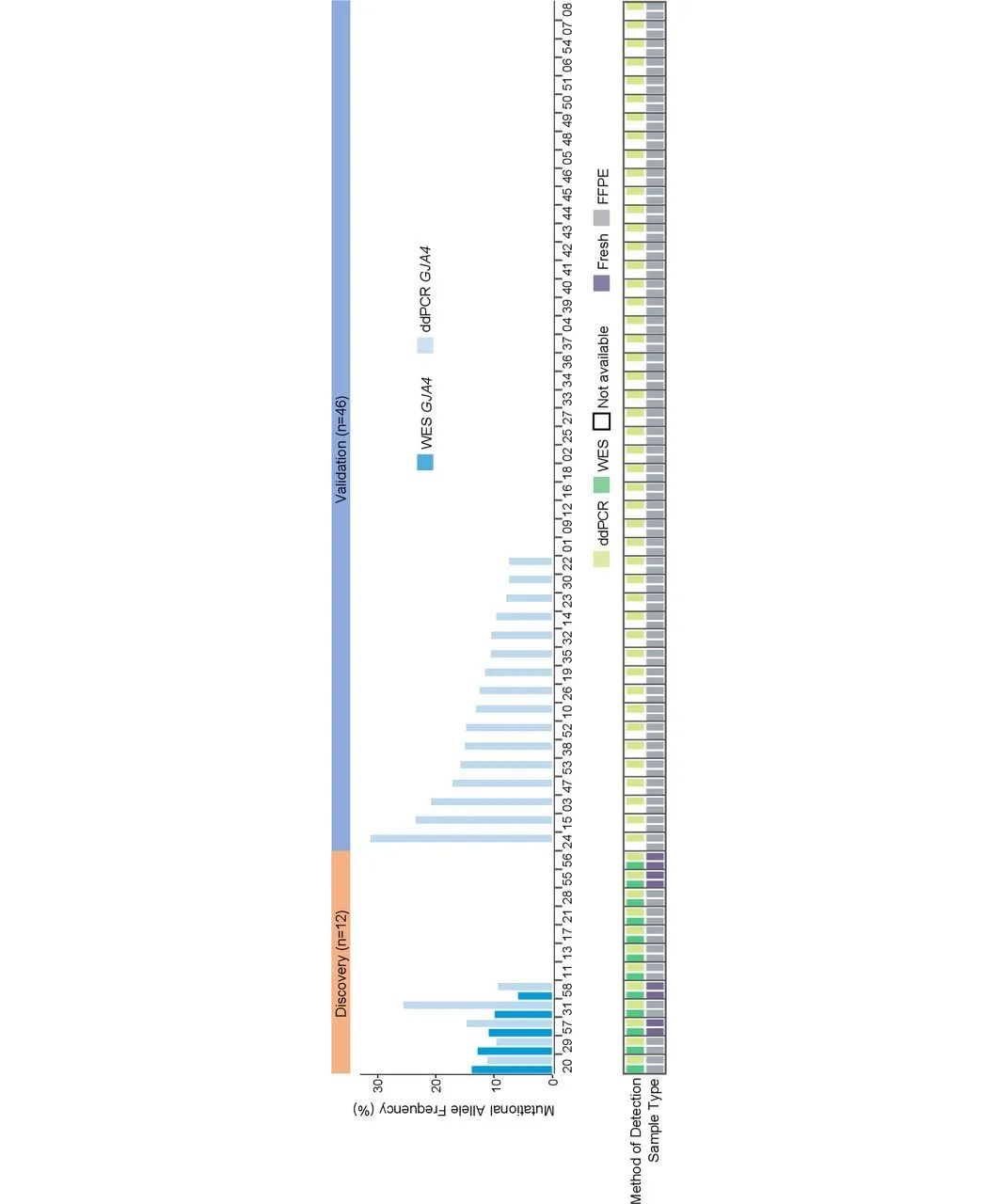
Figure 1. Detection of somatic GJA4 c.121G>T in human ECHs. The top waterfall plot shows the allele frequencies of GJA4 c.121G>T mutation, identified by either the percentage of sequence reads that contained variants on WES or the fractional abundance of variants on ddPCR analysis, in human ECHs. The bottom chart shows details about the samples, including the detected technique, the presence or absence of a paired blood sample, and sample type (fresh-frozen or formalin-fixed, paraffin-embedded tissue). ddPCR, digital droplet PCR; ECH, extra-axial cavernous hemangioma; FFPE, formalin-fixed paraffin-embedded; WES, whole-exome sequencing.
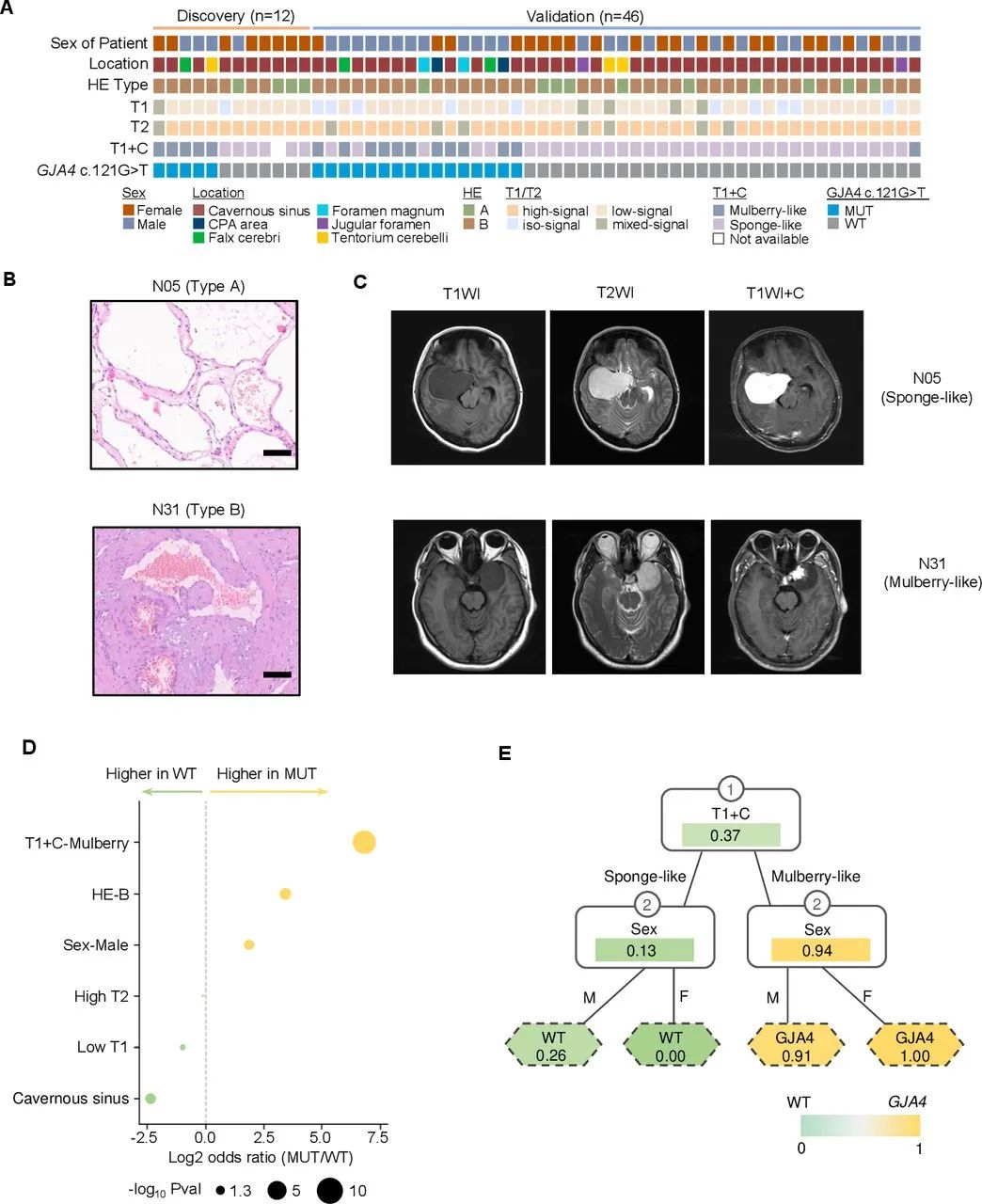
Figure 2. Clinical relevance of somatic GJA4 mutational status. (A) Clinical characteristics of ECHs, including the sex of the patient, ECH location, HE subtypes, MRI-T1WI, T2WI, T1WI+C and the specific GJA4 mutation detected. The patient order is consistent with figure 1. (B) Pathological characteristics of ECH lesions. Scale bar: 100 µm. (C) The MRI characteristics of ECHs. (D) The correlation between clinical features and GJA4 c.121G>T mutation. P value was calculated by Fisher’s exact test. Panel E shows the two-step decision tree model to predict GJA4 c.121G>T mutation. Probability of the mutation was labelled in each node. ECH, extra-axial cavernous hemangioma.
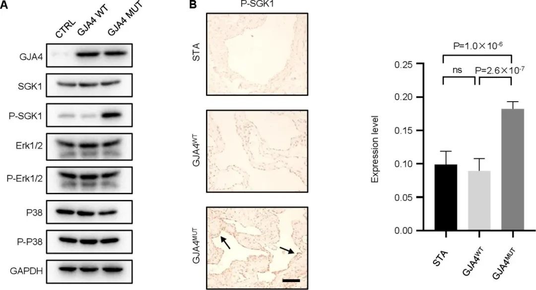
Figure 3. Detection of SGK1 phosphorylation in HUVECS cultures and tissue samples. (A) Immunoblots of HUVECs infected by lentivirus encoding GJA4-WT or GJA4 c.121G>T mutation or empty vector (CTRL) and showed increased phosphorylation of SGK1 but not ERK1/2 or p38. (B) Immunohistochemical staining of a tissue sample of ECH with a GJA4 mutation shows strong staining for SGK1 phosphorylation in endothelial cells lining the vascular lumen (arrows), whereas non-GJA4 mutant sample and superficial temporal artery show little or no staining for SGK1 phosphorylation in endothelial cells (arrowheads). Scale bar: 200 µm. The histogram shows semiquantitative grading of p-SGK1 expression levels in GJA4-WT, GJA4 c.121G>T mutation ECHs and STA. One-way ANOVA followed by Tukey’s multiple comparisons test was used. ANOVA, analysis of variance; ECH, extra-axial cavernous hemangioma; HUVECs, human umbilical vein endothelial cells; STA, superficial temporal artery.
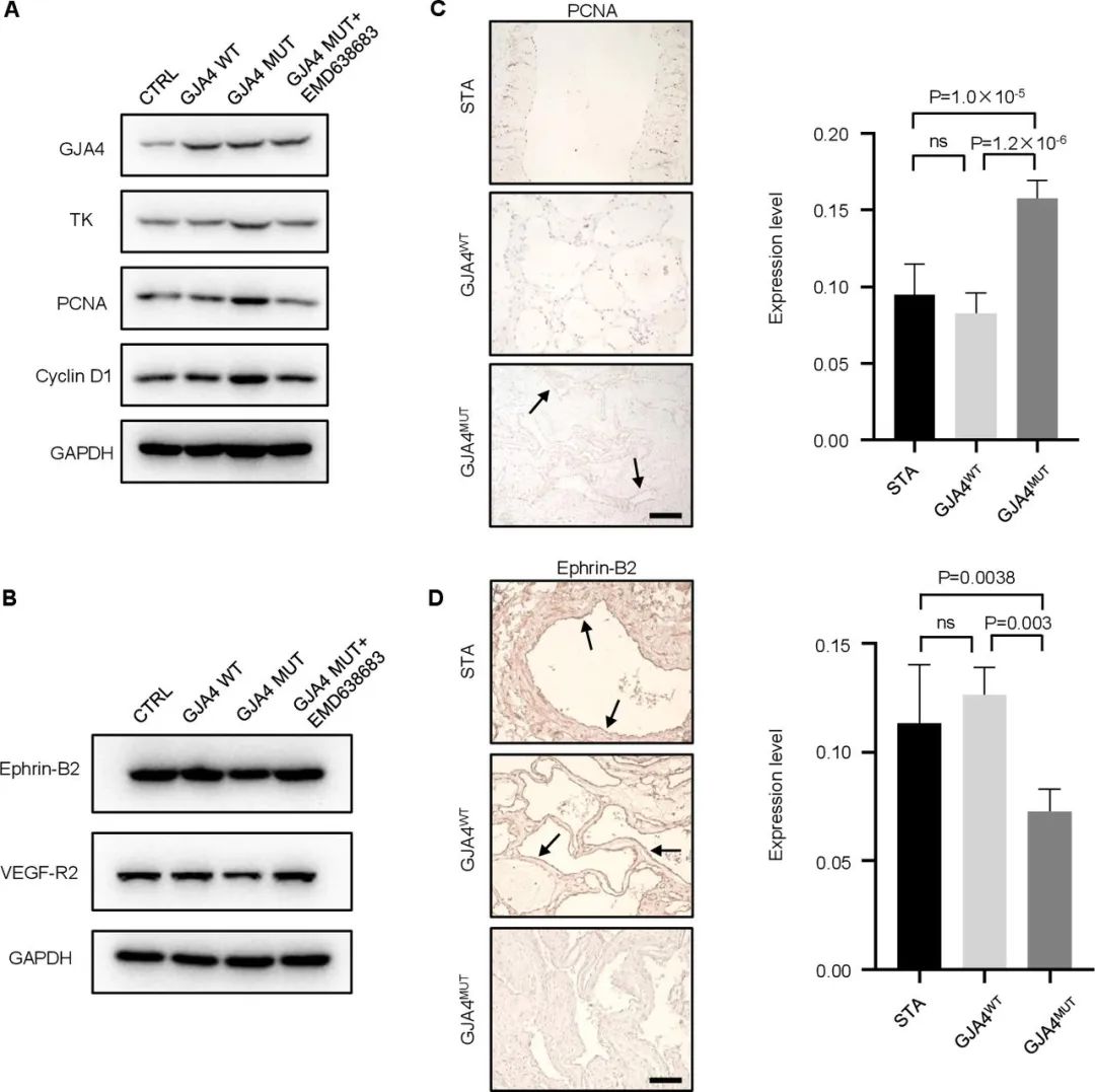
Figure 4. Phenotype of endothelial cells infected with GJA4 c.121G>T mutant lentivirus. (A) Western blot of TK, PCNA and Cyclin D1 in HUVECs infected with CTRL lentivirus, HUVECs overexpressing wild type GJA4, HUVECs overexpressing GJA4 G41C, HUVECs overexpressing GJA4 G41C treated with EMD638683. Results from one representative experiment out of three are shown. (B) Western blot of Ephrin-B2 and VEGF-R2 in HUVECs infected with CTRL lentivirus, HUVECs overexpressing wild type GJA4, HUVECs overexpressing GJA4 G41C, HUVECs overexpressing GJA4 G41C treated with EMD638683. Results from one representative experiment out of three are shown. (C) Immunohistochemical staining of a tissue sample of ECH with a GJA4 mutation shows increased level of PCNA in endothelial cells lining the vascular lumen (arrows), whereas non-GJA4 mutant sample and superficial temporal artery show little or no staining for PCNA in endothelial cells. Scale bar: 200 µm. (D) Immunohistochemical staining of a tissue sample of ECH with a GJA4 mutation shows decreased level of Ephrin-B2 in endothelial cells lining the vascular lumen, whereas non-GJA4 mutant sample and superficial temporal artery show strong staining for Ephrin-B2 in endothelial cells (arrows). Scale bar: 200 µm. One-way ANOVA followed by Tukey’s multiple comparisons test was used. ANOVA, analysis of variance; ECH, extra-axial cavernous hemangioma; HUVECs, human umbilical vein endothelial cells; PCNA, proliferating cell nuclear antigen.
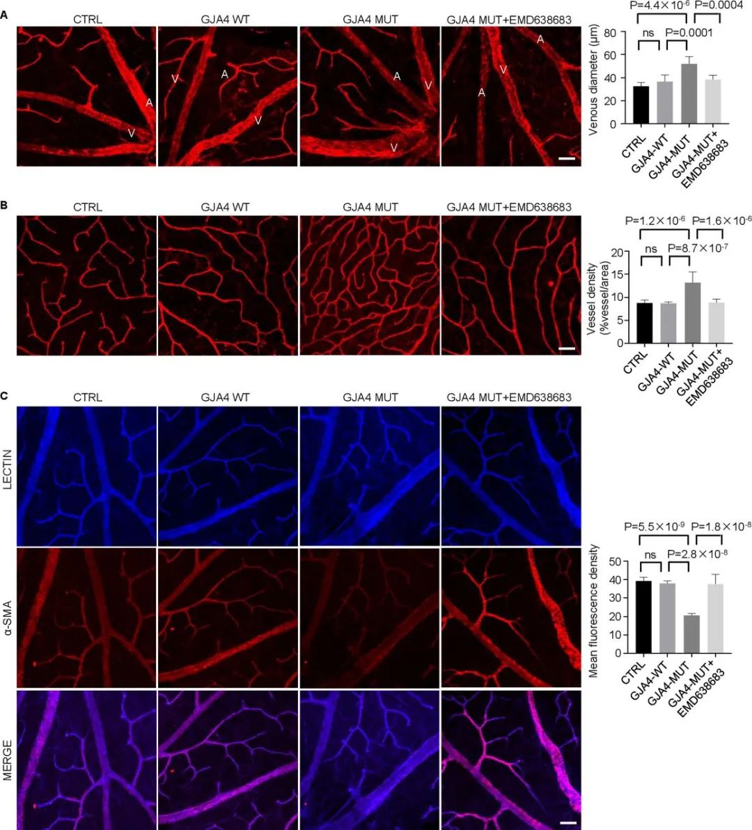
Figure 5. GJA4 mutation regulates retinal angiogenesis in mice. (A) Staining of retinas showed an increase in venous diameter of vessels in the superficial plexuses in the retina of GJA4-mutant mice, compared with GJA4-WT and CTRL mice. EMD638683 reversed the increase of venous diameter caused by GJA4 mutation (n=6). Symbols: A=artery; V=vein. Scale bar: 100 µm. (B) Analysis of Lectin staining (red) at the angiogenic front of C57 mice injected with GJA4-WT, GJA4-MUT and CTRL AAV revealed GJA4 mutation increased the vascular density of vessels, which was reversed by EMD638683 (n=8). Scale bar: 100 µm. (C) Immunostaining showed a reduced α-SMA (red) expression in the vascular networks of retinas of GJA4-mutant mice compared with that observed in GJA4-WT and CTRL mice. Meanwhile, the expression of α-SMA was increased in GJA4-mutant mice treated with EMD638683 (n=3). Scale bar: 100 µm. One-way ANOVA followed by Tukey’s multiple comparisons test was used. ANOVA, analysis of variance.
作者团队明确了一种体细胞GJA4突变,该突变出现在超过三分之一的ECH病变中,并提出ECHs是由于GJA4诱导的脑内皮细胞中SGK1信号通路的激活而导致的血管畸形。

来源:SVN俱乐部
转载已获授权,其他账号转载请联系原账号
神经影像解剖及血供范围分布图-颅脑CT/MR【矢状位】
神经影像解剖及血供范围分布图-颅脑CT【轴位】
查看更多