


200
评论
查看更多
密码过期或已经不安全,请修改密码
修改密码
壹生身份认证协议书
同意
拒绝
同意
拒绝
同意
不同意并跳过


作者:复旦大学脑科学研究院 石子雨
复旦大学脑科学研究院的在读博士石子雨与大家分享于2022年5月23日在线发表于Brain的“Harnessing cortical plasticity via gabapentinoid administration promotes recovery after stroke”一文。
研究背景
探明加巴喷丁类药物GBP的给药是否能通过抑制α2δ2亚基,从而促进损伤的CNS神经元轴突再生,进而在缺血性卒中后促进神经环路功能的恢复。
研究方法
通过玫瑰红腹腔注射和冷光源照射运动皮层的方式构建光血栓性卒中,使用TTC染色,脉管系统标记成像以及激光散斑成像的方式确定光血栓模型的梗死效果和范围。通过使用GFP-M(stock no.007788; RRID: IMSR_JAX 007788)和Aldh1l1-eGFP mice,以及免疫荧光,免疫组化和WB的使用,检测了缺血性梗死后组织的神经元损伤情况,星形胶质细胞变化情况以及α2δ2的表达情况变化。使用Fluoro-Gold逆行标记皮层中的皮质脊髓神经元,检测其中的α2δ2的表达,再使用BDA顺行标记对侧脊髓和脑干内的轴突萌发,来对缺血损伤后的神经轴突再生进行评估。使用了体内多通道记录电生理自发刺激,cFos活性测定激活的神经轴突数量,和行为学来探究小鼠在功能上的恢复情况。通过使用CNO和AAV2-hSyn-hM4D(Gi)-mCherry(1-2e13 GC/ml, 50475, Addgene),对小鼠皮质进行化学基因沉默,对皮质可塑性的增强和功能恢复之间的因果关系进行了验证。
研究结果
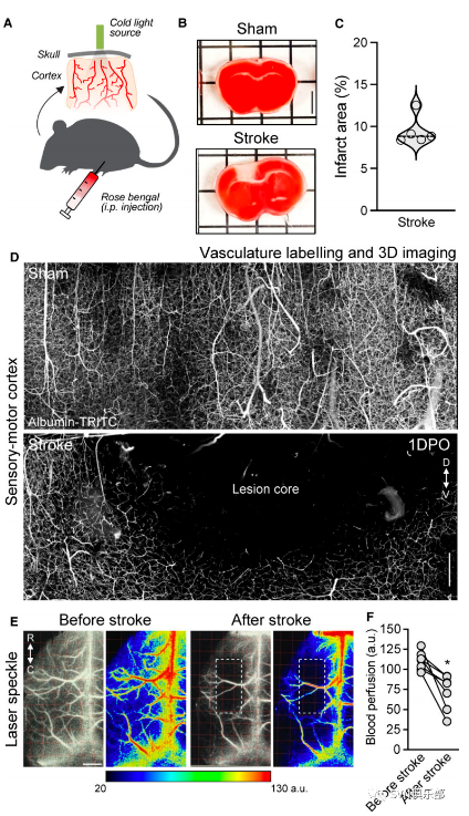
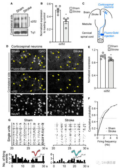
Figure 2. A reduction in α2δ2 expression coincides with changes in electrophysiological properties of corticospinal neurons after stroke.(A)Immunoblot shows α2δ2 expression in the contralateral sensory-motor cortex 7 days after operation. Under reducing conditions, the α2δ2 antibody recognizes two bands at 130 and 105 kDa. Tuj1 is used as the loading control.(B)Quantification of A. Mean and SEM. Biological replicates originated from two independent experiments with sham#1, 2, 3 and stroke#1, 2, 3 from experiment no. 1 and sham#4, 5 and stroke#4 from experiment no. 2. Immunoblots were processed in parallel. Data normalized using loading control(Wilcoxon rank sum test*P < 0.05, sham n = 5 and stroke n = 4).(C)Schematic of retrograde labelling of corticospinal neurons in the brain.(D)Representative fluorescence images of retrogradely labelled corticospinal neurons(arrows)on the contralateral side of the brain 7 days after operation. Scale bar=50 μm.(E)Quantification of D. Mean and SEM(mixed model type Ⅲ test of fixed effects*P < 0.05; sham n = 5 and stroke n = 5; 55-133 neurons/animal, 440-461 neurons/group in total).(F)Differential distribution of firing frequency for all recorded single units(two-sample Kolmogorov-Smirnov test*P < 0.05; sham n = 5 and stroke n = 5; 353-355 single units/experimental group).(G)Raster plots show spontaneous firing within layer V of the sensory-motor cortex 7 days after operation. Bottom: histograms of firing events. Inset: spiking waveform of the single unit; the coloured lines show the average waveform and grey lines show all recorded waveforms.
图3显示了GBP的使用促进了卒中后皮质脊髓的可塑性。
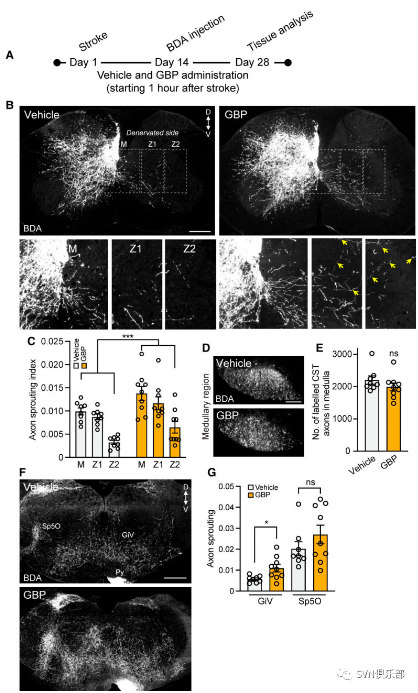 Figure 3. GBP administration promotes corticospinal plasticity after stroke.(A)Experimental scheme of B.(B)Representative fluorescence images of C7 spinal cord sections from adult mice 4 weeks after stroke(D=dorsal, V=ventral). The yellow arrows indicate corticospinal collaterals(bottom). Scale bar=250 μm.(C)Quantification of B. Mean and SEM(type Ⅲ test from linear regression model***P < 0.001; vehicle n = 8 and GBP n = 9).(D)BDA-labelled corticospinal axons in the medullary region. Scale bar=100 μm.(E)Quantification of D. Mean and SEM(Wilcoxon rank sum test; ns=not significant; vehicle n = 8 and GBP n = 9).(F)Representative fluorescence images of brainstem. GiV=gigantocellular reticular nucleus; Sp5O=spinal trigeminal nucleus; Py=pyramidal tract. Scale bar=500 μm.(G)Quantification of F. Mean and SEM(Wilcoxon rank sum test, *P < 0.05, ns=not significant; vehicle n = 8 and GBP n = 9).
图4显示了给予GBP的小鼠有着显著增强的功能连接。
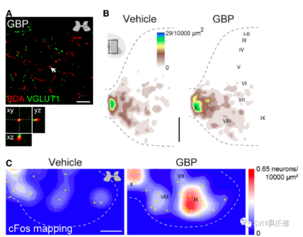
图5表明卒中后1小时开始给小鼠注射GBP可恢复小鼠的前肢功能。
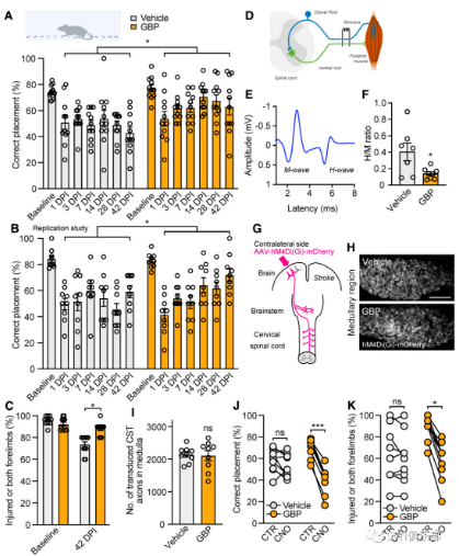
Figure 5. Mice administered GBP beginning 1 h after stroke recover forelimb function.(A)Recovery of forelimb skilled function was assessed using the horizontal ladder rung walking test. Mean and SEM(mixed model with repeated measures using compound symmetry covariance structure and controlled on baseline values*P < 0.05; vehicle n = 11 and GBP n = 11).(B)Replication of the study shown in A. Mean and SEM(mixed model with repeated measures using compound symmetry covariance structure and controlled on baseline values*P < 0.05; vehicle n = 9 and GBP n = 9).(C)Recovery of forelimb symmetry was assessed using the cylinder test.Mean and SEM(mixed model with repeated measures using compound symmetry covariance structure*P < 0.05; vehicle n = 11 and GBP n = 11).(D)Schematic of H-reflex electrophysiological recording.(E)Representative M and H wave responses from forelimb muscles.(F)Hmax/Mmax ratio at 35 days after stroke.Mean and SEM(Wilcoxon rank sum test*P < 0.05; vehicle n = 7 and GBP n = 8).(G)Schematic of chemogenetic silencing.(H)hM4Di(Gi)-mCherry transduced corticospinal axons in the medullary region. Scale bar=100 μm.(I)Quantification of H. Mean and SEM(Wilcoxon rank sum test; ns, not significant; vehicle n = 9 and GBP n = 9). Abrogation of recovery of(J)forelimb skilled walking and(K)forelimb symmetry in rearing after stroke on transient activation of hM4Di(Gi)in corticospinal neurons of mice administered GBP. Aligned dot plot(linear regression model*P < 0.05, ***P < 0.001; ns=not significant; vehicle n = 9 and GBP n = 9).
![]()
研究结论
通过GBP给药的α2δ2药理学阻断是一种易于转化的药理学策略,可促进成年小鼠光血栓性卒中后皮质脊髓通路的可塑性。更重要的是,在给予GBP的小鼠中,大脑对侧完整皮层通路的结构重排推动了小鼠的功能恢复。加巴喷丁类药物在促进急性中枢神经系统创伤后神经功能恢复方面的有益作用正在获得支持。加巴喷丁类药物是临床批准用于治疗多种神经系统疾病的药物,作者的研究结果强调了将GBP重新用作卒中修复的有前途的治疗策略的强大潜力。

Q3:这篇文章中采用了多通道电极阵列记录了皮质第五层神经元的活动,可以看到作者标出了记录电极的位置,那么作者有给予任何刺激吗?如果有是在哪里呢?
A3:作者在设置了记录电极后,主要检测的是小鼠的自发神经活动,所以并没有其他的刺激电极设置。赵恒教授补充,作者对小鼠的前肢进行了机械刺激,来观察神经活动的变化。
Q4:作者使用了光血栓型卒中模型,可以比较一下它和MCAO模型的差异和优劣之处吗?
Q5:请问加巴喷丁类药物和GABA本身有什么样的关系呢?
A5:David Wang教授指出,加巴喷丁在临床上已经使用近20年,主要用于治疗癫痫,是一种GABA类似物,GABA是一种中枢神经系统抑制性神经递质,所以通过加巴喷丁,可以产生类似的效果,抑制中枢神经系统,从而使患者冷静,进而治疗癫痫。然而加巴喷丁背后的机制其实尚不明确,我们发现其实它并非通过模拟GABA来发挥作用,所以它逐渐演变为治疗疼痛的药物。这篇文章发现GBP可以促进对侧的神经再生还是很有意思的。
A6:赵恒教授指出,这篇文章的整体思路比较陈旧,且太过于注重GBP这一药物带来的表型,而并没有对其背后的机制作出更深入的探究,且过于注重神经元上的研究,而现在的研究热点往往需要研究其他神经细胞的共同作用。再者,作者缺少对损伤侧的研究,过于集中在对侧上。
来源:SVN俱乐部

二甲双胍、碘过敏……CT、CTA等检查前需要有哪些注意事项?
徐运:缺血性卒中个体化抗血小板治疗及评估丨CSA&TISC2022
急性脑出血患者收缩压 180 mmHg,紧急降至多少才安全?
ISC 2022|续写新篇章 —— “替奈普酶”精彩继续……
神经影像问答:脑干梗死后华勒氏变性(WD)有什么影像特点和临床表现?
查看更多
 中国医学论坛报
 壹生
壹生 今日肿瘤
今日肿瘤 今日循环
今日循环 今日糖尿病
今日糖尿病 今日口腔
今日口腔 全科周刊
全科周刊 脱贫地区农副产品网络销售平台
京公网安备 11010202008182号
| 互联网新闻信息服务许可证编号:10120190017